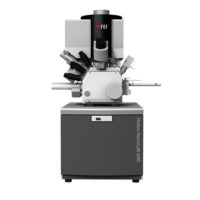
FEI – SEM

scanning electron microscope (SEM) is a type of electron microscope that produces images of a sample by scanning the surface with a focused beam of electrons. The electrons interact with atoms in the sample, producing various signals that contain information about the surface topography and composition of the sample. The electron beam is scanned in a raster scan pattern, and the position of the beam is combined with the intensity of the detected signal to produce an image. In the most common SEM mode, secondary electrons emitted by atoms excited by the electron beam are detected using a secondary electron detector. The number of secondary electrons that can be detected, and thus the signal intensity, depends, among other things, on specimen topography. Some SEMs can achieve resolutions better than 1 nanometer.

Specimens are observed in high vacuum in vacuum or wet conditions in a variable pressure or environmental SEM, and at a wide range of cryogenic or elevated temperatures with specialized instruments.

Description

Easy to use technology

Keep control of experiments with easy to use technology. Reuse jobs and system settings and select multiple regions of interest during job acquisition.

Visualize and navigate during acquisition

Visualize and navigate during acquisition with Thermo Scientific Amira Live Tracker Software to optimize/control your outcome/Automate large 3D volume acquisitions as well as reconstructions.

Protect valuable samples

Protect valuable samples with tested solutions at every acquisition step: debris trap and swipe features ensure sample quality; low vacuum detector enables imaging of highly charged samples.

Easy and fast mounting microtome exchange

Easy and fast mounting microtome exchange for normal SEM operation or automated array tomography with the addition of Thermo Scientific Maps Software.

FEI Microscopic Solution

.